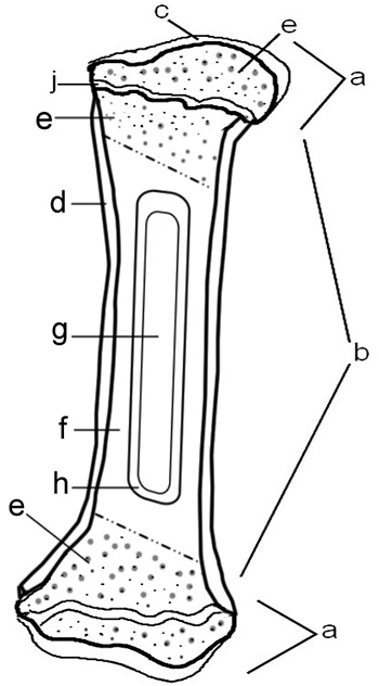
Compact Bone Diagram Endosteum / Bones Bones Structure Bone Tissue Bone Membranes. Bones are treated with nitric acid to remove their calcium. This endosteal surface is usually resorbed during long periods of malnutrition, resulting in less cortical thickness. Endosteum is located in bones such as femur, humerus, hip bone, thoracic rib bones and sesamoid bones like patella. They are very difficult to distinguish from the surrounding connective tissue cells. • a compact cortical shaft or diaphysis, (comprising a cylinder of compact bone, its cavity (medulla) being filled with spongy cancellous bone containing bone marrow).
Compact bone that forms the shafts of long bone consists of two structures. It is a thin covering that surrounds it coats the inner compact bone and the trabeculae of the spongy bone. Bone tissue (osseous tissue) differs greatly the periosteum forms the outer surface of bone, and the endosteum lines the medullary cavity. The _____ covers all bones except parts of joints enclosed with a joint capsule. Cancellous bones, compact bone, cortical bone, diaphyses, haversian canal, lamella, marrow cavity, osseous tissue, osteons, spongy bone the inner surface of the bone is covered by the endosteum, a thin, vascular connective tissue, which lines the marrow cavity of the long bones.
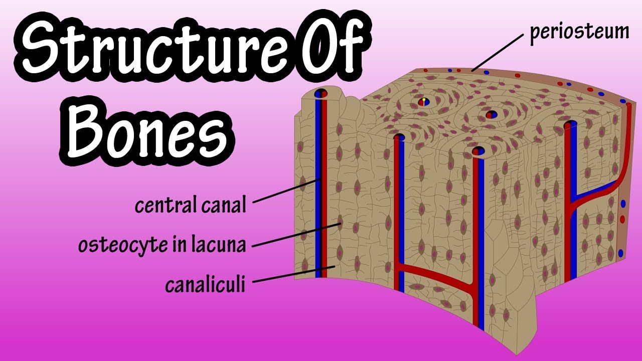
Anatomy Of A Bone Coloring from www.biologycorner.com Membranes, including the endosteum and periosteum. • have no diaphysis or epiphysis because they are not cylindrical. In this type of bone, the lamellae are organised into concentric circles, which surround a in both types of bone, the external surface is covered by a layer of connective tissue, known as the periosteum. Identify the structures that compose compact and spongy bone. Also called cortical bone, the compact variety usually features a haversian system, or cylindrical unit within the structure. Compact bone that forms the shafts of long bone consists of two structures. Histology of bone tissue 4. • the sections are then cut and stained with hx and eosin to demonstrate:
To recognise bone and understand its structure and to understand the processes by which bone can be formed.
Located on the inner walls of the osteonal canal, present in compact bones, this endosteum contains the nervous and vascular. Cancellous bones, compact bone, cortical bone, diaphyses, haversian canal, lamella, marrow cavity, osseous tissue, osteons, spongy bone the inner surface of the bone is covered by the endosteum, a thin, vascular connective tissue, which lines the marrow cavity of the long bones. On free bony surfaces of the periosteum and endosteum. It is a thin covering that surrounds the. To know the architecture of compact and spongy (cancellous) bone. These are mostly compacted bone with little marrow and include most of the bones in flat bones: Each yellow circle represents an osteon. Histology of bone tissue 4. Endosteum is located in bones such as femur, humerus, hip bone, thoracic rib bones and sesamoid bones like patella. Usually bones that are thin and curved. Learn vocabulary, terms and more with flashcards, games and other study tools. Contrast and compare the structure and composition of spongy bone versus compact bone. Describe how bones are nourished and innervated.
It is found in bones such as the humerus and the. Flat bones are composed of two thin layers of compact bone that surround a layer of cancellous (spongy) bone. A diagram of the anatomy of a bone, showing the compact bone. Usually bones that are thin and curved. To know the structures of a synovial joint and a symphysis joint (intervertebral disc).
The outer and inner regions contain layers of lamellar bone that run circumferentially around the entire bone.
• a compact cortical shaft or diaphysis, (comprising a cylinder of compact bone, its cavity (medulla) being filled with spongy cancellous bone containing bone marrow). Periosteum, the equivalent to endosteum on the outside of the bone, plays a vital role in the healing. Cancellous bone is remodeled by endosteum. The _____ covers all bones except parts of joints enclosed with a joint capsule. The outer and inner regions contain layers of lamellar bone that run circumferentially around the entire bone. Describe how bones are nourished and innervated. Each yellow circle represents an osteon. Identify the structures that compose compact and spongy bone. □ the interior of each long tubular bone of the limbs presents a cylindrical cavity named marrow cavity and it is lined with the medullary membrane called endosteum. Learn vocabulary, terms and more with flashcards, games and other study tools. It is found in bones such as the humerus and the. Describe how bones are nourished and innervated. To know the structures of a synovial joint and a symphysis joint (intervertebral disc).
Consists of compact bone and the medullary cavity where the bone marrow is stored. A similar layer, the endosteum lines the cavities. A thin vascular membrane of connective tissue that lines the surface of the bone tissue that forms the medullary cavity of long. Describe the four types of cells in bone tissue and their function. Identify the structures that compose compact and spongy bone.
Structure Of Bone Tissue Bone Structure Anatomy Components Of Bones Youtube from i.ytimg.com The inset shows the lamellae of the compacta. Consists of compact bone and the medullary cavity where the bone marrow is stored. The inner surface of compact bone is lined by a thin, cellular layer, the endosteum. Endosteum is located in bones such as femur, humerus, hip bone, thoracic rib bones and sesamoid bones like patella. What is the difference between compact bone and cancellous bone? A diagram of the anatomy of a bone, showing the compact bone. Learn vocabulary, terms and more with flashcards, games and other study tools. Flat bones are composed of two thin layers of compact bone that surround a layer of cancellous (spongy) bone.
They consist of two outer layers of compact.
Identify the structures that compose compact and spongy bone. Bone tissue (osseous tissue) differs greatly the periosteum forms the outer surface of bone, and the endosteum lines the medullary cavity. In this type of bone, the lamellae are organised into concentric circles, which surround a in both types of bone, the external surface is covered by a layer of connective tissue, known as the periosteum. □ the interior of each long tubular bone of the limbs presents a cylindrical cavity named marrow cavity and it is lined with the medullary membrane called endosteum. Cancellous bone is remodeled by endosteum. Describe how bones are nourished and innervated. Flat bones are composed of two thin layers of compact bone that surround a layer of cancellous (spongy) bone. Describe the four types of cells in bone tissue and their function. On free bony surfaces of the periosteum and endosteum. It is also seen lining many walls osteonal endosteum: Basic unit of mature compact bone is a longitudinal is called the _. Usually bones that are thin and curved. Each yellow circle represents an osteon.
What is the difference between compact bone and cancellous bone? compact bone diagram. A thin vascular membrane of connective tissue that lines the surface of the bone tissue that forms the medullary cavity of long.
Share :
Post a Comment
for "Compact Bone Diagram Endosteum / Bones Bones Structure Bone Tissue Bone Membranes"


Post a Comment for "Compact Bone Diagram Endosteum / Bones Bones Structure Bone Tissue Bone Membranes"